Unpaired MR-CT Brain Image Dataset for Unsupervised Image Translation
The Magnetic Resonance – Computed Tomography (MR-CT) Jordan University Hospital (JUH) dataset has been collected after receiving Institutional Review Board (IRB) approval of the hospital and consent forms have been obtained from all patients. All procedures has been carried out in accordance with The Code of Ethics of the World Medical Association (Declaration of Helsinki).
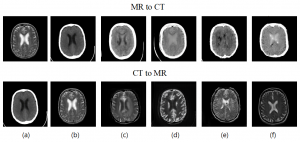
The dataset consists of 2D image slices extracted using the RadiAnt DICOM viewer software. The extracted images are transformed to DICOM image data format with a resolution of 256×256 pixels. There are a total of 179 2D axial image slices referring to 20 patient volumes (90 MR and 89 CT 2D axial image slices). The dataset contains MR and CT brain tumour images with corresponding segmentation masks. The MR images of each patient were acquired with a 5.00mm T Siemens Verio 3T using a T2-weighted without contrast agent, 3 Fat sat pulses (FS), 2500-4000 TR, 20-30 TE, and 90/180 flip angle. The CT images were acquired with Siemens Somatom scanner with 2.46mGY.cm dose length, 130KV voltage, 113-327 mAs tube current, topogram acquisition protocol, 64 dual source, one projection, and slice thickness of 7.0mm. Smooth and sharp filters have been applied to the CT images. The MR scans have a resolution of 0.7×0.6×5 mm3, while the CT scans have a resolution of 0.6×0.6×7 mm3. MR-CT Dataset can be downloaded from here.

*Please note that this data is the property of the Jordan University Hospital and is made available for download for research purposes only. Users are kindly requested to acknowledge the source of this data (https://doi.org/10.1016/j.dib.2022.108109 and implemntation in https://doi.org/10.1016/j.compbiomed.2021.104763) if used for any publication.